Science Highlight: Researchers Uncover Details of Joint Injuries in Children
June 8, 2011
Scientists working in part at the National Synchrotron Light Source (NSLS) have learned new information about how the knee joints of children are damaged as the result of a compression injury, which could happen during sports or play. Their work could help researchers understand how this type of joint injury can progress to secondary osteoarthritis, a condition that occurs due to cartilage loss and causes pain and stiffness.
The research was performed by scientists from the Massachusetts Institute of Technology; Rush University Medical Center, in Chicago; BG Trauma Center Tuebingen and Eberhard Karls University, both in Tuebingen, Germany; and Christian Albrechts University of Kiel, in Kiel, Germany.
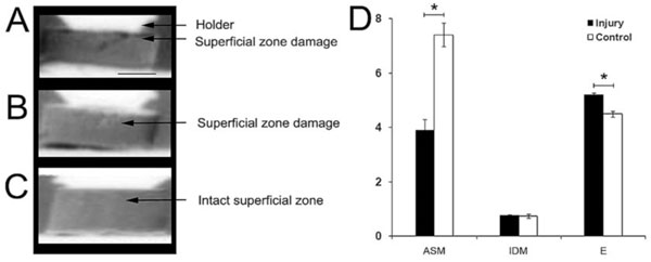
Diffraction-enhanced x-ray imaging (DEI) and texture analysis of injured and control disks of the superficial zone. A-C, Representative DEI images, showing an isolated fissure (A) and slight damage (B) in 2 disks with damage to the superficial zone, as compared with an image of the intact superficial zone of a control disk (C). D, Texture analysis of regions of interest (angular second moment [ASM], inverse difference moment [IDM], and entropy [E]) in the superficial zone of injured and control disks. Bars show the mean ±SEM (n ≥ 5 samples/group). * = P < 0.001.
When an adult experiences a joint compression injury, the type and extent of the damage varies depending on how deep within the cartilage you look. This is due to the inhomogeneous nature of cartilage - at different depths, the composition and related functions are different. But much less is known about compressive injuries in the joints of children.
Here, the scientists used samples from the surface and deeper zones of the joint cartilage of young cows to study compressive injury in detail. The study revealed, as the researchers suspected, that the bulk of the damage was confined to the soft joint surface, where the tissue had been compacted by about 20 percent. This led to the immediate biomechanical failure of the surface.
The group also noticed that even seemingly intact areas had undergone changes in texture, indicating that the structure of the tissue had been altered as a result of the injury.
In the samples from the deeper zones, the study revealed some crimping of the collagen fibers (collagen is the main component of connective tissue), but those samples were otherwise unchanged and biomechanically intact. The fibers were studied using several methods, including an x-ray technique called diffraction enhanced imaging (DEI) that was invented at NSLS in 1995. DEI makes use of how the x-rays diffract within the sample, rather than are absorbed by it, and is very useful in studying soft tissue, as it yields images with more contrast than typical x-ray images.
"DEI allowed us to elucidate changes in injured cartilage samples that other methods did not pick up," said the study's lead author, Bernd Rolauffs. Rolauffs is affiliated with the Massachusetts Institute of Technology, Rush University Medical Center, BG Trauma Center Tuebingen, and Eberhard Karls University.
The researchers also measured the levels of another key connective tissue substance, sulfated glycosaminoglycan (sGAG). Although sGAG content on the surface did not change immediately after injury, levels had decreased by about 18 percent after 48 hours, compared to a loss of just three percent in the deeper zones.
As one of the hallmarks of osteoarthritis development is sGAG loss, these findings could lead to new information on how surface damage can progress to cartilage degradation and ultimately osteoarthritis, a condition that occurs due to cartilage loss and causes pain and stiffness.
This work was funded by the National Institutes of Health and the German Research Foundation.
2011-2429 | INT/EXT | Newsroom