Prototype Breast Cancer Imaging System May Improve Patient Care
Custom-built breast PET-MRI system could improve accuracy of breast cancer imaging, say researchers at SNM’s 56th Annual Meeting
June 15, 2009
The following news release on a new breast cancer imaging system being developed at the U.S. Department of Energy’s Brookhaven National Laboratory was issued by the Society for Nuclear Medicine. The research was conducted by Bosky Ravindranath, a Ph.D. student in the Biomedical Engineering Department of Stony Brook University (SBU), under the direction of Brookhaven Lab Senior Scientist David Schlyer of BNL’s Medical Department, who is also an adjunct professor at SBU. The development of this detector technology was funded by DOE’s Office of Science, Office of Biological and Environmental Research (OBER).
Imaging techniques such as PET and MRI are a direct outgrowth of DOE’s long-standing investment in basic research in chemistry, physics, and nuclear medicine. The ongoing neuroimaging research at Brookhaven is a prime example of how DOE’s national laboratories bring together the expertise of chemists, physicists, and medical scientists to address questions of profound significance for society.
For more information about this research at Brookhaven Lab, contact:
Karen McNulty Walsh, kmcnulty@bnl.gov, 631 344-8350
Mona S. Rowe, mrowe@bnl.gov, 631 344-5056
Society for Nuclear Medicine contact: Amy Shaw, ashaw@snm.org, 703 652-6773
TORONTO—A prototype breast imaging system combining positron emission tomography (PET) and magnetic resonance imaging (MRI) technologies could greatly improve breast cancer imaging capabilities, according to researchers at SNM’s 56th Annual Meeting. Although the system has not yet been tested on humans, initial results from the prototype indicate the system produces a fusion of detailed PET and MRI images that should allow a more accurate classification of lesions in the breast.
“PET and MRI systems are both powerful, noninvasive tools for detecting breast cancer and evaluating treatment, but each of them also has weaknesses,” said Bosky Ravindranath, research assistant working with Dr. David Schlyer at Brookhaven National Laboratory, Upton, N.Y., and lead author of a study on preliminary testing of the prototype. “We believe that combining PET and MRI in a single system will eventually yield highly sensitive and specific breast cancer examinations while at the same time compensating for the shortcomings that exist when using only PET or only MRI.”
Conceptual drawings of the simultaneous PET/MRI breast imaging system being developed at Brookhaven Lab showing the how the PET insert (green) fits inside the MRI scanner and how it will be used with breast cancer patients.
Every year, approximately 180,000 women are newly diagnosed with breast cancer. While the disease is still a leading cause of death among women, breast cancer mortality is declining. In particular, new developments in molecular imaging technologies are dramatically improving the ways in which breast cancer is diagnosed and treated. However, many challenges remain in breast imaging, such as obtaining accurate images of dense breast tissue.
 enlarge
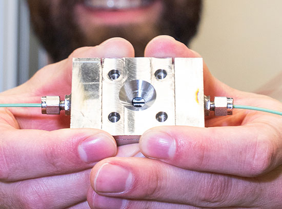
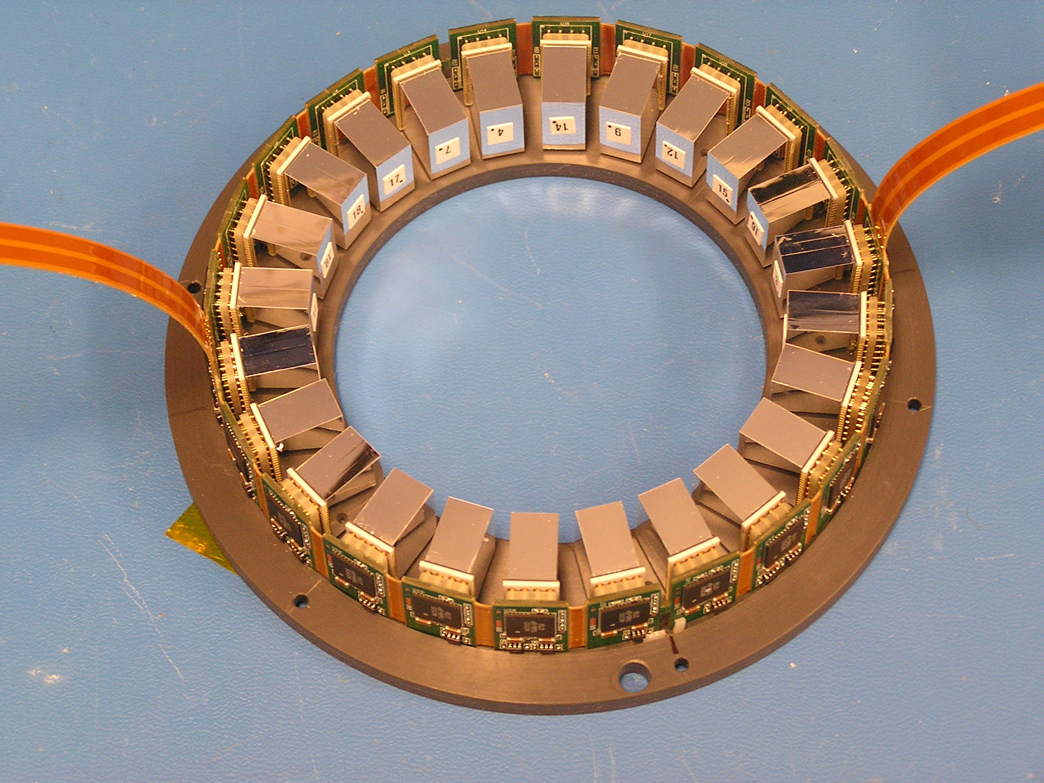
Close up of the PET part of the breast imaging system, showing the individual detector units. This device was developed at Brookhaven Lab in a collaboration of the Medical Department, Physics Department, Instrumentation Division, and Stony Brook University.
When completed, the dedicated breast PET-MRI system will consist of a modular 3D tomographic PET scanner that is inserted inside a dedicated breast MRI coil produced by Aurora Technologies, Inc allowing both PET and MRI images to be taken simultaneously. The modularity of the PET system would allow for the scanner diameter to be adjusted according to patient breast size. Researchers expect the combined modality scanner will provide anatomical information from the MRI to enhance the resolution provided by PET. At the same time, the predictive power of PET in identifying the type of tumor should be able to overcome MRI technology’s traditionally high false-positive rates.
Based on the positive preliminary results, researchers expect to begin testing the system shortly with breast cancer patients.
Scientific Paper 249: B. Ravindranath, S. Junnarker, S.H. Maramraju; S. Southekal, M. Purschke, S. Stoll, D. Tomasi, P. Vaska, C. Woody, D. Schyler, Department of Biomedical Engineering, Stony Brook University, Stony Brook, N.Y.; and Brookhaven National Laboratory, Upton, N.Y. “Initial results from the BNL dedicated simultaneous PET-MRI breast imaging system prototype,” SNM’s 56th Annual Meeting, June 13–17, 2009.
About SNM—Advancing Molecular Imaging and Therapy
SNM is an international scientific and medical organization dedicated to raising public awareness about what molecular imaging is and how it can help provide patients with the best health care possible. SNM members specialize in molecular imaging, a vital element of today’s medical practice that adds an additional dimension to diagnosis, changing the way common and devastating diseases are understood and treated.
SNM’s more than 17,000 members set the standard for molecular imaging and nuclear medicine practice by creating guidelines, sharing information through journals and meetings and leading advocacy on key issues that affect molecular imaging and therapy research and practice. For more information, visit www.snm.org.
2009-10975 | INT/EXT | Newsroom