Unraveling the Origin of Alzheimer's Disease
Researchers find new hints that could explain how the disease spreads in human brains
June 21, 2021
This news release, originally issued by Case Western Reserve University, describes findings that could explain how Alzheimer’s disease spreads in human brains. A team of researchersinvestigated the misfolded proteins that could be the cause for the disease. Among other research methods, the team used the X-ray Footprinting of Biological Materials (XFP) beamline at the National Synchrotron Light Source II (NSLS-II) to study the structure and function of these proteins. The XFP beamline is operated by the Case Western Reserve University Center for Synchrotron Biosciences in partnership with NSLS-II—a U.S. Department of Energy Office of Science User Facility located at Brookhaven National Laboratory. For more information on Brookhaven’s role in this research, please contact Cara Laasch, 631-344-8458, laasch@bnl.gov.
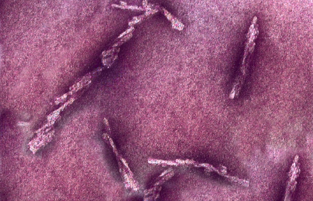
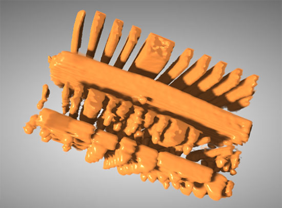
Case Western Reserve University researchers studying prions—misfolded proteins that cause lethal incurable diseases—have identified for the first time surface features of human prions responsible for their replication in the brain.
The ultimate goal of the research is to help design a strategy to stop prion disease in humans—and, ultimately, to translate new approaches to work on Alzheimer’s and other neurodegenerative diseases.
Scientists have yet to discover the exact cause of Alzheimer’s disease, but largely agree that protein issues play a role in its emergence and progression. Alzheimer’s disease afflicts more than 6 million people in the U.S., and the Alzheimer’s Association estimates that their care will cost an estimated $355 billion this year.
Research was done at the Safar Laboratory in the Department of Pathology and the Center for Proteomics and Bioinformatics at Case Western Reserve University School of Medicine, and at Case Western Reserve’s Center for Synchrotron Bioscience at Brookhaven Laboratories in New York. Jiri Safar, professor of pathology, neurology and neurosciences at the Case Western Reserve School of Medicine, leads the work. The report, “Structurally distinct external domains drive replication of major human prions,” was published in the June 17 issue of PLOS Pathogens.
Prions were first discovered in the late 1980s as a protein-containing biological agent that could replicate itself in living cells without nucleic acid. The public health impact of medically transmitted human prion diseases—and also animal transmissions of bovine spongiform encephalopathy (BSE, “mad cow disease”) prions—dramatically accelerated the development of a new scientific concept of self-replicating protein.
Human prions can bind to neighboring normal proteins in the brain, and cause microscopic holes. In essence, they turn brains into sponge-like structures and lead to dementia and death. These discoveries led to the ongoing scientific debate on whether prion-like mechanisms may be involved in the origin and spread of other neurodegenerative disorders in humans.
“Human prion diseases are conceivably the most heterogenous neurodegenerative disorders, and a growing body of research indicates that they are caused by distinct strains of human prions,” Safar said. “However, the structural studies of human prions have lagged behind the recent progress in rodent laboratory prions, in part because of their complex molecular characteristics and prohibitive biosafety requirements necessary for investigating disease which is invariably fatal and has no treatment.”
The researchers developed a new three-step process to study human prions:
- Human brain-derived prions were first exposed to a high-intensity synchrotron X-ray beam. That beam created hydroxyl radical species which, with short bursts of light, selectively and progressively changed the prion’s surface chemical composition. The unique properties of this type of light source include its enormous intensity; it can be millions of times brighter than light from the sun to the Earth.
- The rapid chemical modifications of prions by short bursts of light were monitored with anti-prion antibodies. The antibodies recognize the prion surface features, and mass spectrometry that identifies exact sites of prion-specific, strain-based differences, providing an even more precise description of the prion’s defects.
- Illuminated prions were then allowed to replicate in a test tube. The progressive loss of their replication activity as the synchrotron modifies them helped identify key structural elements responsible for prions’ replication and propagation in the brain.
“The work is a critical first step for identifying sites of structural importance that reflect differences between prions of different diagnosis and aggressiveness,” said Mark Chance, vice dean for research at the School of Medicine and a co-investigator on the work. “Thus, we can now envision designing small molecules to bind to these sites of nucleation and replication and block progression of human prion disease in patients.”
This structural approach, Chance said, also provides a template for how to identify structurally important sites on misfolded proteins in other diseases such as Alzheimer’s, which involves protein propagation from cell to cell in a similar way to prions.
The Safar Laboratory at Case Western Reserve was established in 2008 and focuses on advancing understanding of neurodegenerative diseases, which is crucial for developing new diagnostic and therapeutic strategies. Chance is also director of the Center for Synchrotron Biosciences and the Center for Proteomics and Bioinformatics at the School of Medicine.
The research team used the X-ray beam from the X-ray Footprinting of Biological Materials (XFP) beamline at the National Synchrotron Light Source II (NSLS-II). The XFP beamline is operated by the Case Western Reserve University Center for Synchrotron Biosciences in partnership with NSLS-II—a U.S. Department of Energy Office of Science User Facility.
2021-18963 | INT/EXT | Newsroom


