Visualizing the Internal Structure and Defects in 3D, Self-assembled Nanomaterials
March 31, 2022
 enlarge
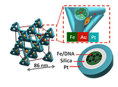
3D assembly and visualization of DNA-guided nanoparticles and frameworks (top). Multielement template (teal) framework of tetrahedra (bottom). Zoomed-in portion shows iron (Fe), silica, and platinum (Pt).
What is the scientific achievement?
A team of scientists from Columbia University, the Center for Functional Nanomaterials, & National Synchrotron Light Source II used advanced X-ray imaging to reveal the structure, composition, and imperfections of 3D nanomaterials designed & synthesized through DNA-mediated assembly and inorganic templating.
Why does this achievement matter?
This work establishes new methods to overcome the difficult challenge of non-destructively verifying the internal structure of complex 3D materials — materials that have promise for catalytic, optical, and energy applications because of their design flexibility.
What are the details?
It is well recognized that a bottom-up nanofabrication is an attractive strategy for arranging functional nano-blocks into 3D materials in order to leverage their functions for novel effects in photonics, biomaterials, electronics, mechanics and other areas. However, the idealized formation of structures has not been achieved. Thus, without a detailed 3D nano-imaging with a single-particle resolution, it is impossible to understand how to tune assembly process and to what degree material performance is affected by imperfections such as defects, dislocations, amorphous regions, and grain boundaries. Currently, the quantitative volumetric assessment of imperfection of nanoscale systems is extremely challenging; this affects both the development of assembly methods and their translation to applications.
The DNA-based assembly offers a tremendous level of control, and it is highly promising as a next-generation nanomaterial fabrication platform. However, the ability to characterize 3D large-scale DNA-assembled nanoparticle architectures at the various assembly states and for different designs are severely limited. This study showcases 3D imaging for the nanoscale nanoparticle lattices assembled with DNA nanostructures. The work also includes a novel type of complex 3D inorganic architectures that combine ordered multimaterial nano-framework, as a continuous phase, and a lattice of discrete nanoparticles, which could be useful for creating materials with novel catalytic, mechanical and electronic properties.
The developed X-ray tomography technique enables the visualization of the designed crystals with unprecedented resolution (7nm) that allows the study of structural imperfection with details never revealed before. It unveils types of defects and their possible origins, paving the way for making perfect artificial structures with novel properties.
CFN Capabilities
- Scanning hard X-ray tomography enabled the 3D visualization of thousands of nanoparticles and motifs at 7-nanometer resolution and with elemental sensitivity
- The CFN Materials Synthesis & Characterization Facility and the NSLS-II HXN, CMS, and SMI beamlines were used in this study
- CMS and SMI beamlines are operated in partnership between CFN and NSLS-II
Publication Reference
A. Michelson, B. Minevich, H. Emamy, X. Huang, Y. S. Chu, H. Yan, O. Gang, “Three-dimensional visualization of nanoparticle lattices and multimaterial frameworks.” Science 376, 6589 (2022).
DOI: 10.1126/science.abk0463
www.science.org/doi/full/10.1126/science.abk0463
Brookhaven Newsroom: Seeing More Deeply into Nanomaterials
Columbia Newsroom: Seeing More Deeply into Nanomaterials
Acknowledgment of Support
We thank the Imaging Facility of CUNY Advanced Science Research Center for instrument use and technical assistance. This research used resources of the Center for Functional nanomaterials and the Hard X-ray Nanoprobe Beamline (HXN) at 3-ID. Small-angle scattering was collected at the Complex Matter Scattering (CMS) instrument at 11-BM and Soft Matter Interfaces (SMI) at 12-ID of the National Synchrotron Light Source II, which are part of the US Department of Energy, Office of Science facilities at Brookhaven National Laboratory under contract DE-SC0012704.
2022-19603 | INT/EXT | Newsroom