MeV-STEM Shows Promise in Advanced Imaging of Thicker Biological Samples
April 9, 2025
 enlarge
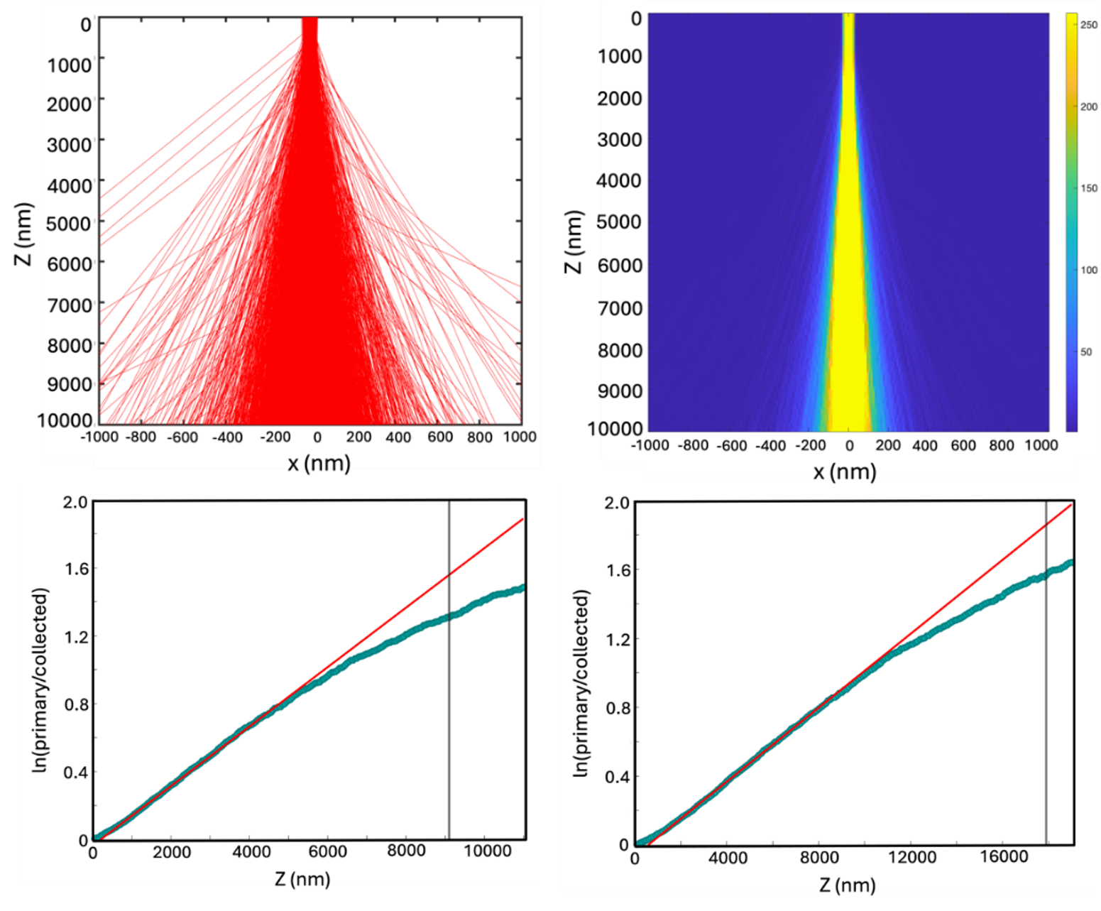
Electron beam trajectory and intensity profiles at 3 MeV. (A) Trajectories of 10,000 electrons in amorphous carbon. (B) The heatmap plot reveals beam intensity. (C) Relationship between logarithmic ratio of intensities and thickness in amorphous carbon (D) and amorphous ice. Vertical lines indicate the maximum linear depth.
The Science
Computer simulations demonstrate mega-electron-volt scanning transmission electron microscopy (MeV-STEM) as a promising way to image samples up to 10 micrometers thick.
The Impact
Imaging thicker samples with nanoscale resolution paves the way for new insights into biology and semiconductor research.
Summary
Cryo-electron microscopy (cryo-EM) is an excellent technique for imaging biological specimens at the nanometer scale but faces challenges when dealing with thicker samples. Researchers need a method to study samples up to 10 micrometers thick while preserving fine structural details. To address this, scientists at the Laboratory for BioMolecular Structure, part of the U.S. Department of Energy’s (DOE) Brookhaven National Laboratory, are exploring megaelectron-volt scanning transmission electron microscopy (MeV-STEM), which employs high-energy electrons capable of penetrating thicker samples with minimal degradation in resolution.
In this study, researchers conducted computer simulations to analyze how high-energy electrons interact with different materials. They examined variables such as electron energy, material composition, and detector configurations to assess how effectively electrons traverse various samples. The simulations revealed that electron beam spread and intensity are influenced by both the atomic number and material density. While elements with higher atomic numbers scatter electrons more strongly, materials with high density, like carbon, also induce significant scattering due to their abundance of atoms compared with ice, where biological samples are embedded. This explains why carbon, despite having a lower atomic number than oxygen, exhibited greater beam broadening in the simulations.
Furthermore, the study found that focusing the electron beam deeper within the sample, rather than at the top surface, resulted in a narrower, more precise beam profile, particularly at higher convergence semi-angles. This insight is valuable because maintaining small convergence angles is challenging in laboratory settings, making defocus depth a critical parameter for optimizing image quality.
The results also confirmed that electron intensity follows the Beer–Lambert law, which describes how light or electrons decrease in intensity as they pass through material. Notably, at 3 megaelectron-volts (MeV), electrons can penetrate significantly deeper compared to lower energies. For instance, in silicon dioxide, the penetration depth at 3 MeV is approximately six times greater than at 300 kiloelectron-volts (0.3 MeV). In amorphous ice, the effect is even more pronounced, with penetration reaching 11.5 times deeper.
These findings advance the potential of MeV-STEM for imaging thicker biological and semiconductor samples, paving the way for more detailed and accurate structural studies in diverse scientific fields.
Download the research summary slide (PDF)
Related Links
Contact
Liguo Wang
Brookhaven National Laboratory
lwang1@bnl.gov
Xi Yang
Brookhaven National Laboratory
xiyang@bnl.gov
Publications
Quintard, B., Yang, X., & Wang, L. (2025). Quantitative Modeling of High-Energy Electron Scattering in Thick Samples Using Monte Carlo Techniques. Applied Sciences, 15(2), 565. https://doi.org/10.3390/app15020565
Funding
This project was partially supported by the U.S. Department of Energy, Office of Science, Office of Workforce Development for Teachers and Scientists (WDTS) under the Science Undergraduate Laboratory Internships Program (SULI) and partially funded by the DOE Office of Biological and Environmental Research, grant number KP1607011.
2025-22479 | INT/EXT | Newsroom