Summary of Intramural NIH/ Brookhaven National Laboratory Cell Phone Study in JAMA
Cell phones show effect on brain activity most pronounced near the antenna; finding is of unknown clinical significance
February 23, 2011
In a study of the effects of cell phone usage on brain cell activity, NIH scientists and their colleagues at the U.S. Department of Energy’s Brookhaven National Laboratory found that 50 minutes of cell phone usage (with the phone muted to avoid confounding effects from auditory stimulation) elevated brain glucose metabolism significantly in the parts of the brain closest to the phone’s antenna. Elevations in glucose metabolism, a measure of brain cell activity, were correlated with the estimated strength of the electromagnetic field emitted by the phone in those regions. The findings are published in the in the February 22, 2011, issue of JAMA.
“Although we cannot determine the clinical significance, our results give evidence that the human brain is sensitive to the effects of radiofrequency-electromagnetic fields from acute cell phone exposures,” said Nora Volkow, the study’s lead author.
Discrepancies among studies on the effects of RF-electromagnetic fields (RF-EMF) from cell phones on the human brain highlight the need for additional research. Prior studies of the acute effects of cell phone use on human brain function, including measurements of cerebral blood flow monitored by positron emission tomography (PET), have yielded inconsistent results, which might have reflected, in part, the small sample sizes of such studies (the largest studies had 14 subjects).
 enlarge
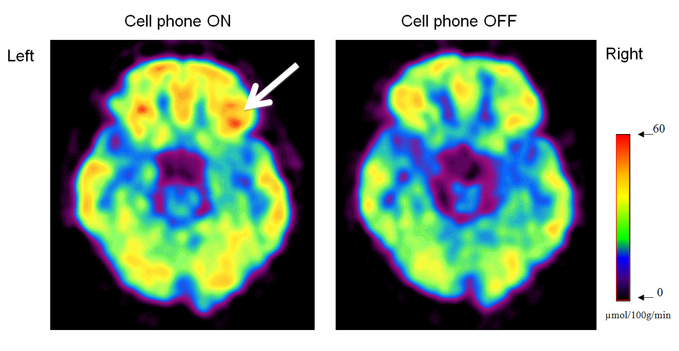
Arrow in the left image shows the location in the orbitofrontal cortex in one subject where glucose metabolism was increased during cell phone use. Red and orange areas shows higher brain metabolic activity. On the right is a baseline image with the cell phone turned off, showing lower activity.
The current study, also using PET, provides a more direct measure of brain activity than cerebral blood flow. It uses a radioactively “tagged” form of glucose known as [18F]fluoro-deoxyglucose, or FDG, to measure glucose metabolism in specific regions of the brain.
“FDG is a direct substitute for glucose, the brain’s fuel. Measuring its concentration in the brain gives a highly specific measure of brain cell metabolism, which is a more direct measure of brain activity than measures of blood flow,” Volkow said.
Also, because brain glucose metabolic measures obtained with FDG reflect the averaged brain activity occurring over a 30-minute period, this method can assess the cumulative effects of cell phone exposure on resting brain metabolism, unlike blood flow measures, which isolate a more restricted point in time.
Scientists conducted the study in 47 healthy individuals. All participants had two scans done on separate days. On both days, prior to the scans, two cell phones, one placed on the left and one on the right ear, were used so subjects wouldn’t know which cell phone was active. For one of the days both cell phones were off. For the other day the right cell phone was on but muted while receiving a call from a recorded text. The order of conditions was randomly assigned, and participants did not know when an active phone was being tested.
With the cell phones secured to their heads, the subjects sat in a quiet room, not speaking, with eyes open for 20 minutes prior to being injected with the FDG tracer, and then for 30 more minutes, for a total cell phone exposure time of 50 minutes . RF-EMF emissions were recorded once before the call (background) and every five minutes during the “on” phase to ensure that the call was not terminated. Using computational and photographic methods, the scientists were also able to calculate the relative amplitude of the cell phone’s electric field at every position in the brain.
After the exposure period, the phones were removed and the subjects were placed in the PET scanner for measurements of brain activity.
Results
There were no differences in overall brain metabolism between the on and off conditions, but during the on condition, the specific regions of the brain closest to the phone’s antenna showed significant increases in brain glucose metabolism. The regions expected to have the greater absorption of RF-EMF from the cell phone exposure were the ones that showed the largest increases in glucose metabolism.
“The linear association between cell phone-related increases in metabolism and electric field strength suggests that the metabolic increases are secondary to the absorption of RF-EMF from cell phone exposures,” Volkow said. “Further studies are needed to assess if the effects we observed could have potential long-term consequences.”
This research was funded by the Intramural Research Program of the National Institutes of Health (NIH) using infrastructure supported at Brookhaven Lab by DOE’s Office of Science.
For further information or to speak with Nora Volkow, please contact the NIH Press Office: 301-496-5787
2011-11239 | INT/EXT | Newsroom