Reflecting Antiferromagnetic Arrangements
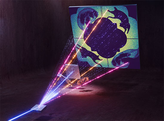
Scientists reflected extremely coherent x-rays of antiferromagnetic domains to make them visible
January 31, 2019
 enlarge
Inactive Akt1 can be activated by two mechanisms. In one mechanism, phosphorylated serine 473 interacts with the linker between the kinase and PH domain. In the other, phosphorylated serinve 477 and Threonine 479 result in the displacement of the PH domain. Image courtesy of Cell 174(4):897-907 (2018).
The Science
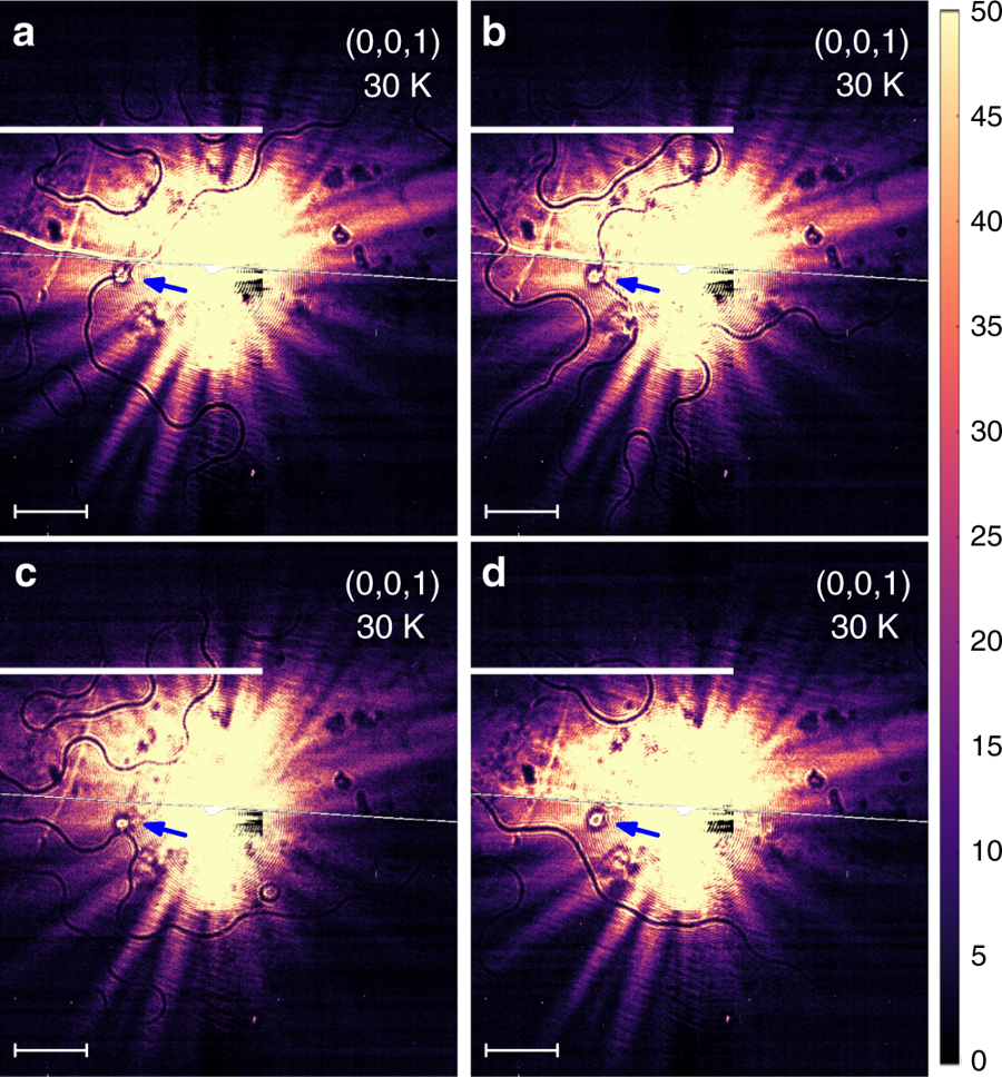
Scientists imaged antiferromagnetic (AFM) domains using a new coherent x-ray diffraction technique and observed evidence of the interaction between domain walls and structural defects in the material.
The Impact
The capability to visualize and manipulate magnetic domains plays an essential role in the development of novel spintronic devices. Recent advances in antiferromagnetic spintronics have given rise to novel concepts for developing electronic devices.
Summary
A team of scientists has demonstrated an x-ray imaging technique that could enable the development of smaller, faster, and more robust electronics. This technique, called coherent resonant magnetic x-ray diffraction, addresses a primary limitation in the emerging research field of “spintronics,” or spin electronics, which involves magnetic materials known as antiferromagnets (AFMs). This limitation is the inability to image antiphase magnetic domains. In all magnetic materials, there are distinct regions—magnetic domains—in which the electron spins are arranged in a regular manner. For AFMs the net magnetization of the material is zero, making domain imaging challenging.
The electronic properties of domain walls can be different from those in the bulk of the material, and scientists can use this as an advantage to find and, hopefully, manipulate the domains by external perturbations.
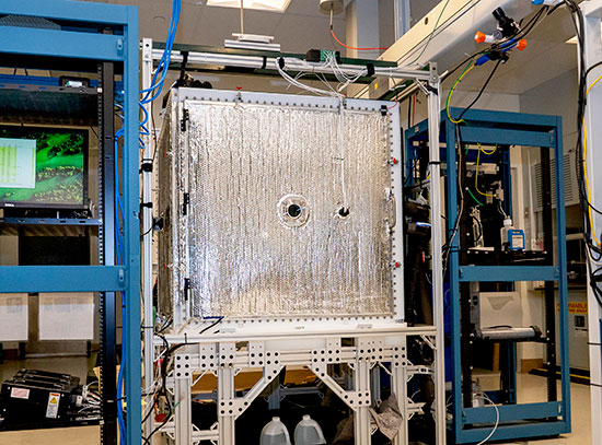
The team used the Coherent Soft X-ray Scattering (CSX) beamline at the National Synchrotron Light Source II (NSLS-II), a U.S. Department of Energy (DOE) Office of Science User Facility at DOE’s Brookhaven National Laboratory, to image the domain walls of the collinear antiferromagnet Fe2Mo3O8 during thermal treatment. The scientists demonstrated the capability to visualize antiphase AFM domain boundaries with five micrometer spatial resolution and in a 0.3 × 0.3 millimeter area. The whole measurement took less than one second and does not require any additional numerical algorithms.
The speed of the technique makes it ideally suited for dynamic experiments and the scientists were able to study how the magnetic domains changed in real time as they heated the sample to “melt” (remove) its antiferromagnetic order and cooled it to bring back the order in the form of the domain arrangement.
They discovered that some of the domains were free to move with each thermal cycle while others were not.
Download the research summary slide
Related Links
Brookhaven Lab Press Release: “Reflecting Antiferromagnetic Arrangements”
Contact
V. Kiryukhin
Rutgers University, Piscataway
vkir@physics.rutgers.edu
Publications
M. G. Kim, H. Miao, B. Gao, S.-W. Cheong, C. Mazzoli, A. Barbour, W. Hu, S. B. Wilkins, I. K. Robinson, M. P. M. Dean, V. Kiryukhin, “Imaging antiferromagnetic antiphase domain boundaries using magnetic Bragg diffraction phase contrast“ Nature Communication 9, 5013 (2018). DOI: 10.1038/s41467-018-07350-3
Funding
Work by the Rutgers group, including sample growth and X-ray scattering, was supported by the U.S. Department of Energy (DOE) under Grant No. DOE: DE-FG02-07ER46382. This research used resources at the 23- ID-1 beamline of the National Synchrotron Light Source II, a DOE Office of Science User Facility operated for the DOE Office of Science by Brookhaven National Laboratory under Contract No. DE-SC0012704. Work in the Condensed Matter Physics and Materials Science Division was supported by the DOE Office of Science, Office of Basic Energy Sciences, under Contract No. DE-SC00112704. M.P.M.D. acknowledges support by the DOE, Office of Basic Energy Sciences, Early Career Award Program under Award No. 1047478.
2019-17484 | INT/EXT | Newsroom