Chemical Imaging with Unprecedented Nanoscale Resolution
Scientists at NSLS-II showed the chemical interplay in batteries with unprecedented resolution, opening a pathway to new research opportunities
November 30, 2020
 enlarge
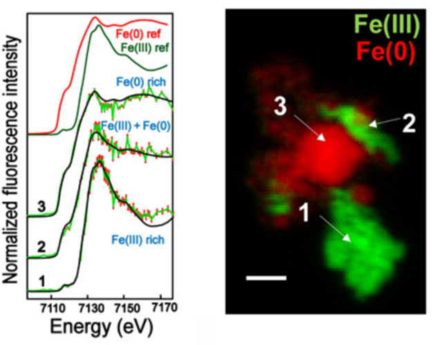
This image shows how different iron species are distributed within one battery particle using high resolution x-ray imaging at the nanoscale. Image credit: A. Pattammattel, et. al. Sci. Adv. 6 : 37, eabb3615 (2020)
The Science
Scientists demonstrated spectroscopic chemical imaging at unprecedented nanoscale resolution.
The Impact
Understanding chemical interplay at the nanoscale in materials such as batteries and microchips allows for the faster development of new technologies; this work demonstrates the ability to resolve the chemical structure of such materials with high sensitivity and <50 nm spatial resolution.
Summary
From solar cells to microchips, our modern life is becoming increasingly reliant on various nanotechnologies. This ever-growing field of applications comes with a high demand for research tools that can unravel a holistic view of a material’s chemical and physical properties. While researchers have access to a myriad of microscopy tools at different length scales to study various aspects of nanomaterials, there remains a gap when it comes to resolving the chemical speciation, with high resolution, within thick samples.
In this work, scientists report nanoscale chemical speciation by combining two hard x-ray techniques: a scanning nanoprobe and fluorescence-yield x-ray absorption near-edge structure (nano-XANES) spectroscopy. The combination of these two techniques offers a new pathway to study the chemical make-up of materials with high sensitivity and resolution.
The team demonstrated the resolving power of nano-XANES by mapping two different iron states of a reference sample composed of stainless steel and hematite nanoparticles with 50 nm scanning steps. Following this demonstration, the team used the new technique to study secondary phases in lithium iron phosphate (LFP) particles of lithium-ion batteries. They found individual iron-phosphide nanoparticles in pristine LFP, whereas partially (de)lithiated particles showed iron-phosphide nanonetworks. These findings shed light on the contradictory reports on iron-phosphide morphology in the previously published literature.
The team used the Hard X-ray Nanoprobe (HXN) beamline at the National Synchrotron Light Source II (NSLS-II) to enable these nano-XANES studies. NSLS-II is a U.S. Department of Energy (DOE) Office of Science User facility located at DOE’s Brookhaven National Laboratory that offers access to a wide range of multimodal and multidisciplinary materials characterization tools. The researchers also verified their results using two other imaging beamlines, the Full Field X-ray Imaging (FXI) beamline and the X-ray Fluorescence Microprobe (XFM) beamline, at NSLS-II.
Both studies demonstrate the advanced capabilities of nano-XANES, bridging the gap between the current capabilities of spectromicroscopy methods, and providing exciting research opportunities across multiple disciplines.
Download the research summary slide
Contact
Hanfei Yan
Brookhaven National Laboratory
hyan@bnl.gov
Publication
A. Pattammattel, R. Tappero, M. Ge, Y. S. Chu, X. Huang, Y. Gao, H. Yan, High-sensitivity nanoscale chemical imaging with hard x-ray nano-XANES, Science Advances 6 : no. 37, eabb3615 (2020). DOI: 10.1126/sciadv.abb3615
Funding
This research used 3ID, 18ID, and 4BM beamlines of the National Synchrotron Light Source II, a U.S. Department of Energy (DOE) Office of Science User Facility operated for the DOE Office of Science by Brookhaven National Laboratory under contract no. DE-SC0012704.
2020-18792 | INT/EXT | Newsroom