X-ray Imaging Technique Highly Sensitive to Trace Impurities in a Crystal
August 30, 2024
 enlarge
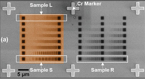
Scanning electron microscopy images show the silicon samples implanted with gallium ions (darker regions). The grayscale gradient is due, in part, to minor height differences caused by sputtered silicon atoms. (Credit: Small Methods May 1:e2301610 (2024)).
The Science
Scientists used X-rays to detect tiny clusters of gallium atoms implanted within a pure silicon crystal.
The Impact
This level of detection sensitivity could help scientists harness the potential optoelectronic and quantum properties of impurities/dopants, laying a foundation for new technologies.
Summary
As they work to develop new quantum technologies, scientists often study incredibly small structures, including single atomic impurities in insulators that can act as quantum sensors and single photon sources. To study these structures, scientists need imaging methods that are incredibly sensitive and won't damage the samples.
X-ray fluorescence (XRF) microscopy has proven to be a powerful tool to provide nanoscale imaging with chemical specificity, but the limits of its sensitivity were not known. While there are other methods that can detect smaller amounts of material, such as energy-dispersive X-ray spectroscopy (EDX) and Kelvin probe force microscopy (KPFM), XRF offers the advantage of being able to identify nanoscale-sized clusters with elemental sensitivity buried within a sample without damaging it. In a recent study led by the University of Surrey, scientists pushed the limits of the powerful XRF technique at the Hard X-ray Nanoprobe (HXN) beamline at the National Synchrotron Light Source II (NSLS-II), a U.S. Department of Energy (DOE) Office of Science User Facility at DOE’s Brookhaven National Laboratory, to investigate implanted gallium impurities in silicon.
Samples were made by implanting gallium into a silicon crystal using a focused ion beam. The team controlled factors like beam current, spot size, and pulse duration very precisely. Gallium atoms have a large mass difference in comparison to silicon, providing good contrast between the XRF photon energies of the impurity material and the host material. The researchers found that they could detect incredibly small features containing just 650 gallium atoms! By using calibration samples with known numbers of atoms, they could accurately measure and map these tiny features. This study suggests that with planned upgrades to light source facilities, we might soon be able to detect the features of single atoms with this technique in the near future.
Download the research summary slide (PDF)
Contact
Benedict N. Murdin
University of Surrey
b.murdin@surrey.ac.uk
Publications
Masteghin, M.G., Gervais, T., Clowes, S.K., Cox, D.C., Zelyk, V., Pattammattel, A., Chu, Y.S., Kolev, N., Stock, T.J.Z., Curson, N.J., Evans, P.G., Stuckelberger, M., Murdin, B.N., "Benchmarking of X-Ray Fluorescence Microscopy with Ion Beam Implanted Samples Showing Detection Sensitivity of Hundreds of Atoms.” Small Methods May 1:e2301610 (2024). DOI: 10.1002/smtd.202301610
Funding
M.G.M. and S.K.C. acknowledge financial support from the Engineering and Physical Sciences Research Council (EPSRC) [Grant No. EP/X018989/1]. T.G. and V.Z. acknowledge support from EPSRC [DTP, Grant No. EP/T518050/1]. BNM is also grateful for EPSRC support [Grant No. EP/N015215/1]. ATI-based authors would like to thank the UK National Ion Beam Centre [EPSRC, Grant No. EP/X015491/1] and UCL-based authors were financially supported by EPSRC [Grant No. EP/W000520/1, EP/R034540/1 and EP/V027700/1]. N.K. thanks the EPSRC Centre for Doctoral Training in Advanced Characterization of Materials [Grant No. EP/S023259/1]. P.G.E. acknowledges support from the US Department of Energy Office of Basic Energy Sciences through contract DE-FG02-04ER46147. E.D.X. analysis was supported by EPSRC [Grant No. EP/V036327/1].
2024-22070 | INT/EXT | Newsroom


