
Multimodal imaging from the micro- to nanoscale of elemental abundances and chemical speciation in a wide range of heterogeous natural materials
The Bioimaging Program at Brookhaven Lab's National Synchrotron Light Source II (NSLS-II) offers a wide range of imaging and spectroscopic microscopy tools for the structural and chemical analysis of natural materials from the micro- to the nanoscale. The beamlines in this program specialize in the study of natural materials in plant, environmental, planetary, and sustainability sciences.
Program Beamlines
X-ray Fluorescence Microprobe
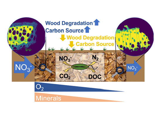
The XFM beamline offers scientists unique and versatile tools for characterizing elemental abundances and chemical speciation in heterogeneous materials. XFM spans a broad energy range and is designed for multimodal and mesoscale imaging, as well as spatially-resolved spectroscopy in diverse scientific fields, including catalysis and energy sciences as well as biology, environmental sciences and energy sciences.
Quantitative Cellular Tomography
The QCT beamline will provide the researchers with a high-throughput analytical approach of imaging frozen hydrated intact cells. Equipped with a cryo-light microscope, the beamline will also have built-in capability of correlation on subcellular morphology and functionality. QCT supports a wide variety of research areas including molecular environmental science, plant and soil sciences, earth and planetary sciences, and life and biological sciences.
Life Science X-ray Scattering
The LiX beamline supports scanning mapping and tomographic imaging of biological tissues, based on both scattering and fluorescence contrasts. Our user research covers a wide range of topics, including engineered plants for bioenergy applications, environmental management, biomedical research, and bio-inspired materials.
Frontier Synchrotron Infrared Spectroscopy
Researchers use the FIS beamline to probe the structure and behavior of materials under extreme conditions. FIS can replicate the high temperatures and pressures found deep within planets, enabling studies of material properties and reactions using infrared electronic and vibrational spectroscopy. Under ambient conditions, FIS also offers infrared spectromicroscopy and imaging for spatially resolved research in biogeochemistry, Earth, and environmental sciences.
Bioimaging News
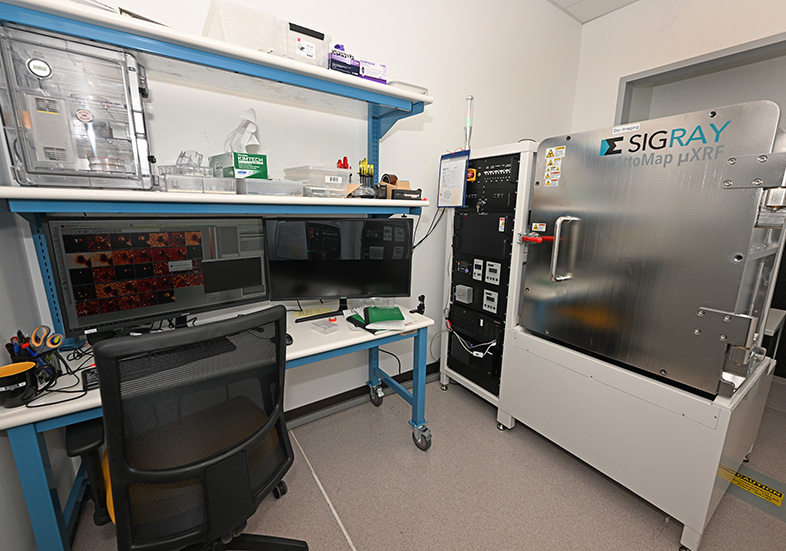
BioImaging Core (BIC) of NSLS-II/CBMS is responsible for an offline X-ray Fluorescence Imaging station called AttoMap-310 (Sigray Inc.). AttoMap covers a broad range of energies (5 source targets) for sensitivity to both light (eg P) and heavy elements (eg U) with spatial resolutions ranging from 5 to 15 microns.
Tiffany Victor is a bioimaging scientist that uses synchrotron imaging techniques to address questions in biological and environmental science, with an emphasis on metal and metalloprotein distribution in cells and tissues and the inorganic N distribution in plant-fungi symbiotic systems.
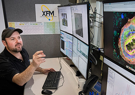
Ryan Tappero, soil chemist and the lead beamline scientist at the X-ray Fluorescence Microscopy (XFM) beamline, captures an image of a manganese nodule from an agricultural soil prepared as petrographic thin section.
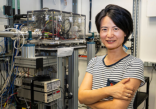
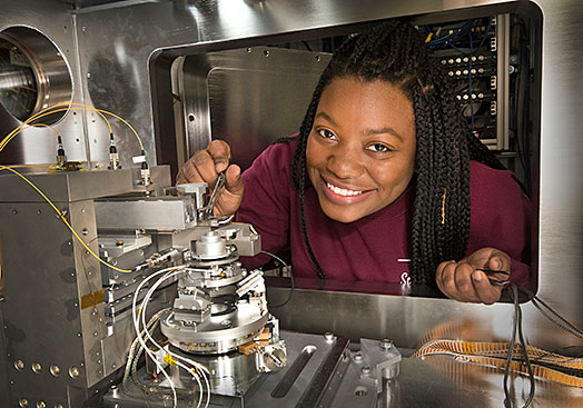
Yang Yang is an X-ray nano-imaging scientist with expertise in 2D and 3D X-ray nano-imaging with synchrotron-based techniques and leads the development of the upcoming Quantitative Cellular Tomography (QCT) beamline.
Our Partners
Synchrotron Earth and Environmental Science (SEES) is a NSF-funding consortium whose mission is to advance research and education in synchrotron-based Earth and environmental science to better understand our planet from the atmosphere to the core. SEES support the XFM, XPD, and FIS beamlines at NSLS-II.