One-Minute Nanotomography
NSLS-II's Full Field X-ray Imaging beamline can image samples in 3-D faster than ever before
September 30, 2018
 enlarge
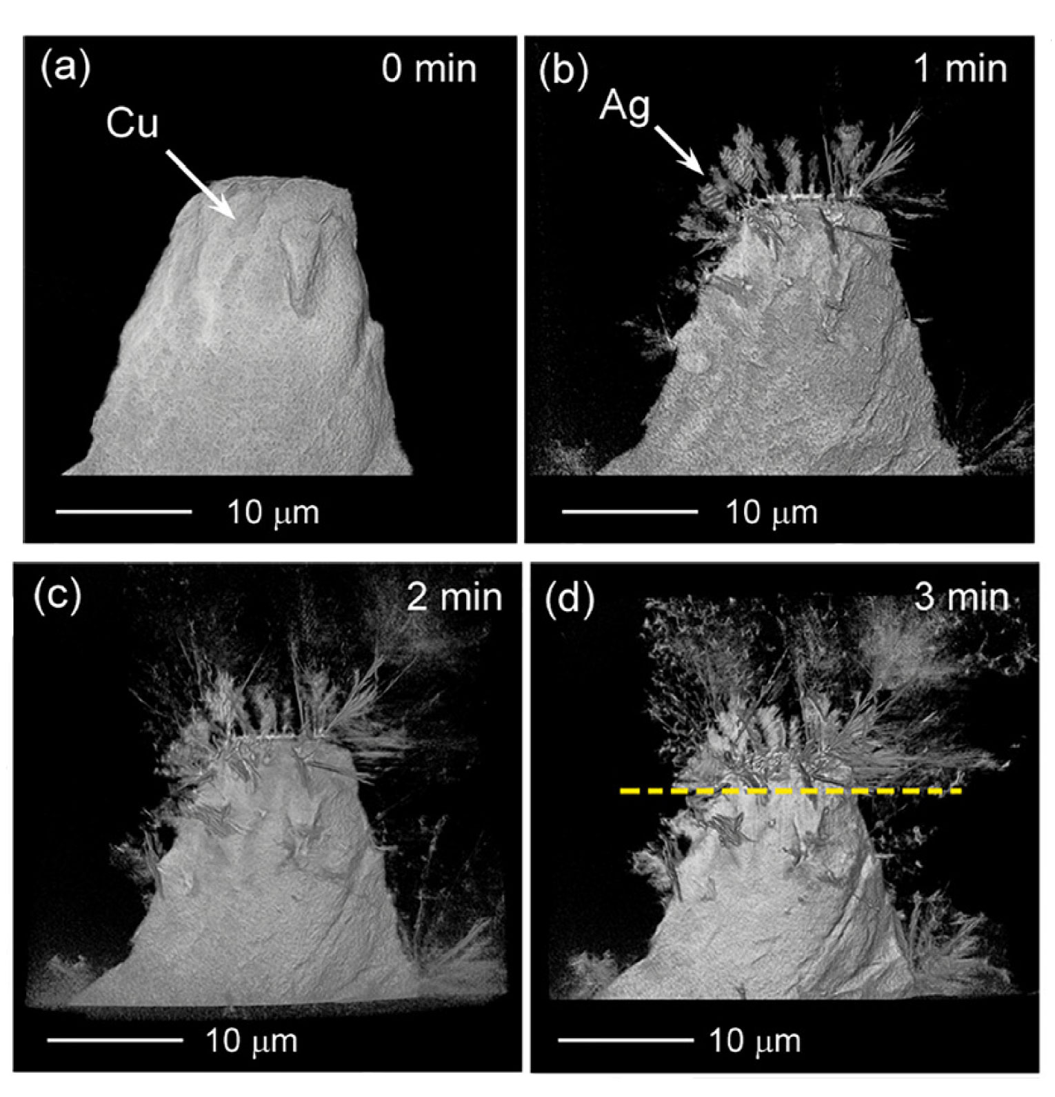
The top left image shows the bare cupper (Cu) base before the growth of the silver (Ag) whiskers begins. The images b-d show the different stages of whisker growths in one-minute increments. Image credit: Appl. Phys. Lett. 113, 083109 (2018)
The Science
Scientists achieved 3-D nanotomography with a sub-50-nanometer resolution in one minute.
The Impact
This accomplishment demonstrates that synchrotron x-ray nanotomography can be used for imaging fast changing systems, such as structural morphology evolution during chemical reactions, in operando, and in real-time measurements.
Summary
At synchrotron light sources, scientists can use transmission x-ray microscopes (TXM) to see into samples with much higher resolution than optical microscopes can provide, revealing extraordinary details. TXM is a powerful technique for non-destructive 3-D imaging with nanometer-scale spatial resolution for samples ranging from biological cells to energy storage materials. However, the typical acquisition time with a hard x-ray TXM at a light source is longer than 10 minutes for one 3-D nanotomography dataset with sub-50nm spatial resolution. This significantly limits the types of 3-D dynamics and fast changes in materials that can be investigated using this technique
In this study, scientists demonstrated the first one-minute 3-D nanotomography with a sub-50-nanometer resolution using full field transmission x-ray microscopy. This achievement was made possible with an in-house-designed and newly commissioned TXM instrument at the Full-field X-ray Imaging (FXI) beamline at the National Synchrotron Light Source II (NSLS-II). NSLS-II is a U.S. Department of Energy (DOE) Office of Science User Facility located at DOE’s Brookhaven National Laboratory.
This capability represents an order of magnitude decrease in the time required for studying sample dynamics with nanometer spatial resolution. To test the new instrument, the scientists measured a number of test samples, including the growth of silver whiskers on copper, a process that is much faster than usual nanotomography could capture.
The scientists from the FXI beamline are looking forward to working with researchers from all around the world to uncover yet-to-be-seen processes using this new tool.
Related Links
Feature Story: Making X-ray Microscopy 10 Times Faster
Contact
Wah-Keat Lee
Brookhaven National Laboratory
wklee@bnl.gov
Publication
M. Ge, D. S. Coburn, E. Nazaretski, W. Xu, K. Gofron, H. Xu, Z. Yin, and W. K. Lee. One-minute nano-tomography using hard X-ray full-field transmission microscope. Applied Physics Letters 113, 083109 (2018). DOI: 10.1063/1.5048378
Funding
This research used resources at FXI beamline (18-ID) of the National Synchrotron Light Source-II, a U.S. Department of Energy (DOE) Office of Science User Facility operated for the DOE Office of Science by Brookhaven National Laboratory under Contract No. DE-SC0012704. This research used resources of the Center for Functional Nanomaterials, which is a U.S. DOE Office of Science Facility, at Brookhaven National Laboratory under Contract No. DE-SC0012704.
2018-17470 | INT/EXT | Newsroom