Phase Contrast X-ray Imaging of Bio-samples by Single-shot Picosecond ICS
To capture a high resolution single shot X-ray image of a live object, four parameters are especially important – short- duration pulses, small transverse size of the source, high protonflux detectable by realistic imaging techniques, and mono-chromaticity. All these are offered by the laser driven Thomson source. Another important feature is partial coherency that helps enhance the resolution of low contrast details of a semi-transparent object placed into the beam by detecting the phase shift, together with the absorption of X-rays. Phase-contrast imaging is especially important when the samples to be visualized are weakly absorbing (low Z). Hence, biological and clinical studies, such as angiography and mammography, benefit from phasesensitive imaging techniques. The PI and co-workers demonstrated earlier that a laser synchrotron source(s) can support phase-contrast imaging. The ATF experiment was the first to achieve this with a Thomson X-ray source. To illustrate potential biological application of this technique so enabled, we present in the figure below a single shot image of a wasp taken with a 1-ps exposure time at a distance 1 m from it. In particular, the details of the borders of low absorption are visible due to the phase-contrast effect. This method will be useful for quantitative computed tomography applications of Thomson sources.
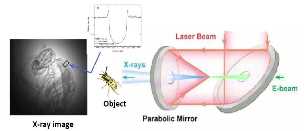
High-resolution radiographic image of a wasp acquired with a single one picosecond shot of the ATF’s Thomson source. The insert illustrates edge enhancement due to interference fringes emerging around the border of areas with different refraction index (phasecontrast effect).
Oliva, P., et al., Quantitative evaluation of single shot inline phase contrast imaging using an inverse compton x-ray source. Applied Physics Letters, 2010. 97(13): p.134104.