Brookhaven and the Nobel Prize
2009
Nobel Prize in Chemistry
Venkatraman Ramakrishnan, of the Medical Research Council Laboratory of Molecular Biology in Cambridge, UK, a former employee in Brookhaven’s biology department, and Thomas A. Steitz of Yale University shared the prize with Ada E. Yonath of the Weizmann Institute of Science for studying the structure and function of the ribosome.

The Structure and Function of the Ribosome
Two of the three recipients of the 2009 Nobel Prize in Chemistry conducted a substantial part of their award-winning research at Brookhaven’s National Synchrotron Light Source (NSLS). They used the NSLS and other synchrotron sources to produce atomic-level images that helped reveal the inner-workings of the ribosome, a cellular complex responsible for producing the thousands of proteins that are required for living cells. Venkatraman Ramakrishnan, of the Medical Research Council Laboratory of Molecular Biology in Cambridge, UK, a former employee in Brookhaven’s biology department and a long-time user of the NSLS, and Thomas A. Steitz of Yale University, also a long-time NSLS user, shared the prize with Ada E. Yonath of the Weizmann Institute of Science.
The ribosome translates the genetic instructions encoded by DNA into chains of amino acids that make up proteins. It is composed of two subunits: 30S, which reads the code, and 50S, which links up the amino acids.
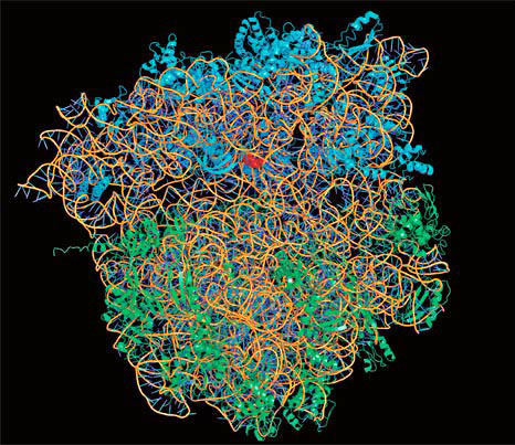
An x-ray structure of a bacterium ribosome. Credit: the Royal Swedish Academy of Sciences
Starting in the late 1990s, both Ramakrishnan and Steitz used a technique called x-ray crystallography at the NSLS to gather atomic-level structures of these two ribosome subunits, Ramakrishnan on 30S and Steitz on 50S. In this technique, scientists analyze how a beam of powerful x-rays is scattered by molecules arranged in a crystal to determine the positions of the molecule’s individual atoms.
Crystallography data from the NSLS contributed directly to these scientists’ prizes, along with research conducted at the Advanced Photon Source (APS) at Argonne National Laboratory and the European Synchrotron Radiation Facility.
The structures of 30S and 50S have been crucial to understanding everything from how the ribosome achieves its amazing precision to how different antibiotics bind to the ribosome, knowledge that could help researchers come to grips with the problem of multi-drug-resistant bacteria.
Determining the structure of the ribosome made it possible for Ramakrishnan and his colleagues to image antibiotics bound to the ribosome, leading to a better understanding of their action, which could help in the development of novel drugs. In this 2004 talk, Ramakrishnan conveys the excitement in the field that has arisen as a result of these discoveries.