Ultra-high Spatial Resolution Meets Multimodal Imaging
The HXN beamline at Brookhaven Lab's National Synchrotron Light Source II now offers a combination of world-leading spatial resolution and multimodal imaging
May 31, 2018
 enlarge
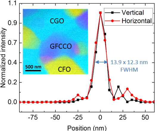
Using a nano-focused x-ray beam, researchers were able to image an emergent phase of an ionic membrane (inset). This incredibly small x-ray beam was created by specially designed multi-layer Laue lenses (MLLs) that were manufactured at Brookhaven Lab. Image credit: Nano Future 2, 011001, (2018)
The Science
Scientists achieved multimodal imaging with ultra-high resolution (~12 nm) and will use it to study novel and complex materials in detail.
The Impact
This is a major milestone for hard x-ray microscopy that will enable new breakthroughs in applied materials science and related fields.
Summary
By channeling the intensity of x-rays, synchrotron light sources can reveal the atomic structures of countless materials. From proteins to batteries, scientists can study the chemistry, structure, and elemental distribution of materials using various techniques at light sources like the National Synchrotron Light Source II (NSLS-II), a U.S. Department of Energy (DOE) Office of Science User Facility located at DOE’S Brookhaven National Laboratory. However, many of these techniques are limited by what resolution they can achieve.
Scientists at the Hard X-ray Nanoprobe (HXN) beamline at NSLS-II have demonstrated the beamline’s ability to observe materials down to 12 nanometers and can now combine this high spatial resolution with multimodal scanning. In order to realize this ambitious goal, the scientists needed to address many technical challenges, such as reducing environmental vibrations, developing effective characterization methods, and perfecting the optics. A key component for the success of this project was developing a special focusing optic, a multilayer Laue lens (MLL).
In this work, a team of researchers used HXN’s unique multimodal imaging capabilities to investigate a mixed ionic-electronic conducting ceramic-based membrane material employed in solid oxide fuel cells and membrane separations (compound of Ce0.8Gd0.2O2−x and CoFe2O4), which revealed the existence of an emergent material phase and quantified the chemical complexity at the nanoscale.
Combining multimodal and high-resolution imaging is unique and makes NSLS-II the first facility to offer this capability in the hard x-ray energy range to visiting scientists. The achievement will present a broad range of applications.
Download the research summary slide
Related Links
Feature Story: New Capabilities at NSLS-II Set to Advance Materials Science
Contact
Hanfei Yan
Brookhaven National Laboratory
hyan@bnl.gov
Publication
H. Yan, N. Bouet, J. Zhou, X. Huang, E. Nazaretski, W. Xu, A. Cocco, W. Chiu, K. Brinkman, Y. Chu. Multimodal hard x-ray imaging with resolution approaching 10 nm for studies in material science. Nano Future 2, 011001, (2018). DOI: 10.1088/2399-1984/aab25d
Funding
This research used resources of the Hard X-ray Nanoprobe (HXN) Beamline 3-ID of the National Synchrotron Light Source II (NSLS-II), a U.S. Department of Energy (DOE) Office of Science User Facility operated for the DOE Office of Science by Brookhaven National Laboratory under Contract No. DE-SC0012704. Additional financial support came from an Energy Frontier Research Center on Science Based Nano-Structure Design and Synthesis of Heterogeneous Functional Materials for Energy Systems (HeteroFoaM Center) funded by the US Department of Energy, Office of Science, Office of Basic Energy Sciences (Award no. DE-SC0001061).
2018-17479 | INT/EXT | Newsroom