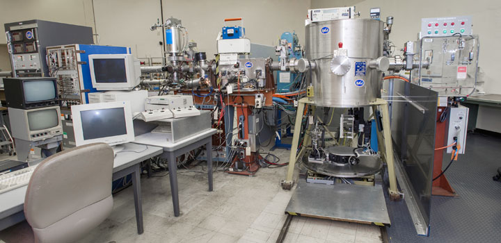
Single Event Upset Test Facility
Introduction
The uninterrupted and progressive miniaturization of microelectronic devices while resulting in more powerful computers, has also made these computers more susceptible to the effects of ionizing radiation. This is of particular concern for space applications due to the radiation fields encountered outside the protective terrestrial atmosphere and magnetosphere. Starting in 1987, a coalition of US government agencies (NSA, NASA, NRL and USASSDC ) collaborated with BNL to develop a powerful and user-friendly test facility for investigating space-radiation effects on micro-electronic devices[1]. The main type of effects studied are the so called Single Event Upsets (SEUs) where ionization caused by the passage of a single heavy ion produces a bit flip and hence a computer error or a system crash. Such events can also lead to destructive latchups and burnouts which must be prevented at almost any cost. The wide variety of beams available at the BNL Tandem, while lower in energy than the average heavy ions encountered in space, do cover the entire range of specific ionization and of linear energy transfer values (LET), and are therefore well suited for simulating these effects under controlled conditions. To make this possible, new systems had to be developed to produce uniform beams and to precisely control and monitor ion fluxes ranging from about 100 to about 10,000,000 particles per cm2 per second. This facility has evolved into a highly automated and well interlocked computer controlled system which can be easily operated by first-time users on the day of their arrival. High beam purity, good beam uniformity and precise dosimetry are provided to a large community of American and European users in support of their space programs.
The single event upset test facility
The heavy ions used for SEU testing are generated using an upgraded model MP tandem Van de Graaff accelerator [2] which is operated at a maximum terminal voltage of 15 MV. The range of energies and corresponding LET values achievable for a few representative beams are shown in fig.1. Many other ions are available and, up to date, about 35 different elements have been accelerated.
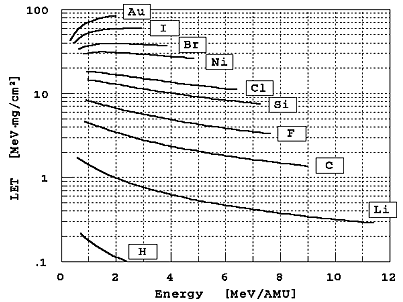
Fig. 1. Beam energies and corresponding LET values in silicon for a few representative beams available at the Brookhaven Single Event Upset Test Facility. Many other beams are also available. More than 35 different elements have been accelerated.
You can now calculate LETs on line. Most common TVDG ion species and a variety of targets are available. The program accepts energies at or well beyond those available at the TVDG.
Negative ions are generated in a sputter-type ion source operated at ~-150 kV with respect to ground and a first mass selection is performed by means of an inflection magnet. This magnet which has a mass resolution of 1/40 is operated at ~-120 kV and is provided with a hall-probe gaussmeter. Then the ~150 keV negative ions are injected into the accelerator contained in a pressure vessel which is filled with insulating gas containing ~50% SF6, ~40% N, and ~10% CO2, pressurized to about 10 atm. The negative ions are accelerated in an evacuated acceleration tube to the high voltage terminal which can be operated at voltages ranging from ~1 to 15 MV. In the high voltage terminal the negative ions are converted to positive ions by allowing them to pass through a carbon foil which causes several electrons to be stripped off the ions but which is thin enough (typically ~5 ug/cm2) to cause an almost negligible energy loss. A second stripper foil, located at the 75% voltage point can be optionally used to achieve even higher charge states and consequently higher energy gain during the last stage of acceleration.
A beam-calibrated generating voltmeter located at the wall of the pressure vessel is used to measure the terminal voltage with a precision of 0.1-0.2%. The energy of the different charge-state components emerging from the accelerator is therefore also known to this precision. A highly precise double-focusing magnet used in conjunction with narrow object and image slits is then used to select one of these components and to determine the energy with even greater precision. The field in this magnet is measured by means of a nuclear magnetic resonance probe and the magnetic rigidity of the selected component is thus determined with a resolution of ~10-3 and a precision of ~10-4. In spite of this high resolution, there are occasions in which different charge-state components of some ions can have close enough magnetic rigidities as to cause possible confusion or beam contamination, especially when both strippers are used. A cross-field velocity selector (Wien filter) has been installed to further purify the beam and a number of procedures have been implemented to detect and eliminate any residual contaminants.
First a computer search is performed to identify possible known beam contaminants. Charge-state combinations which may generate beams of similar rigidities are then avoided. This computer search can be skipped in the case of well established beams for which extensive experience exists. The next step is to select at least three different charge states by varying the terminal voltage. The measured voltages and beam intensities are then compared with computer predictions and the energies are verified by means of a silicon detector (see below). If no anomalies are encountered the desired beam is deflected in the switcher magnet and is allowed to traverse the multiple scattering foil. Some of the ions scattered by this foil enter a silicon surface barrier-detector, the pulse height spectrum of which is recorded in a multichannel analyzer. Here the energy and purity of the beam are checked making use of the fact that if different charge-state components are present they will have different energies. If a single peak is observed at the correct position the final energy is calculated by making a small correction to take into account the energy loss in the gold foil, and the beam is sent into the target room. Here an independent energy and purity check is performed with one of several alpha-particle calibrated silicon surface barrier detector systems. Some of these detectors are also partly covered with aluminum foils of different, calibrated thicknesses and can thus be used for precise on-line LET measurements.
The ion flux reaching the part being tested can be adjusted by utilizing ion source controls, a variable aperture at the accelerator entrance, object and image slit settings, scattering foil thickness selection, and beam sweeping amplitude changes. A method useful for reducing the flux to very low values consists of using a carbon stripping foil at the image location to generate a charge state distribution and then selecting a peak component of this distribution with the switcher magnet. Doing this allows one to easily maintain a beam intensity at the regulating slits sufficient for stable accelerator operation.
Beam uniformity at the device mounted on the goniometer stage is achieved by multiple scattering in the foil at the entrance to the target room by two dimensional sweeping with two steerer magnets or by a combination of these methods. Different sweeping frequencies in the range 5-30 Hz are selected for each magnet and their lack of synchronization is monitored with an oscilloscope. The uniformity is monitored and verified by a set of four plastic scintillation detectors surrounding the useful part of the beam and by a fifth detector which can be rotated into the central on-axis position. These five detectors are provided with precisely machined collimators of identical area. Usually the counting rate in the central detector is somewhat higher than the average of the other four and a corresponding automatic correction is applied to the fluence as measured by the outer detectors. The divergence of the beam while traveling from the plane of the detectors to the location of the tested device is taken into account through a small, calculated geometric correction.
During a prolonged measurement, the central detector is periodically and automatically rotated into the beam, the correction factor is updated and if it becomes too large the users and the operators are alerted. The flux and uniformity are constantly monitored in the diagnostic chamber and displayed numerically and graphically in a specially designed PC based system [3] which is located in the control room and which also provides alarms when the flux or other parameters fall outside selected limits.
The beam can be intercepted by a Faraday cup and by a "parts shutter". The beam is collimated by a variable and rotatable iris which is adjusted so as to avoid bombarding parts adjacent to the one under study. By maintaining the plane of the iris parallel to the test board the illuminated area on the board remains constant when both planes are rotated. For some tests, other than normal incidence is chosen to vary the "effective" LET value.
The entire target-room system is computer controlled and automated with user-friendly menu-driven software. The system can "remember" and reproduce the positions of a large number of devices on the board. The information is fed into the system by positioning each device with the aid of a joy-stick and by pressing the "enter" key. To aid in the location of the devices a light beam, which is almost collinear with the ion beam, is generated with a laser and reflected by a mirror. The collimated light spot on the device is then observed on a monitor connected to a television camera. Once the test board information has been entered, a test sequence can be set up by selecting desired fluences and effective LET values. After recording the number of upsets for different LET values the system provides on-line display of the single event upset cross-section curves and documentation in the form of plots, printed tables and information recorded on floppy discs. Good examples of such data can be found in ref. [4].
If the chamber needs to be opened the total vacuum cycle time is less than 5 minutes. The large number of chips that can be simultaneously installed and the fast, totally automated vacuum system add to the unprecedented testing efficiency which is achieved with this system.
References
- [1]
- P. Thieberger, E.G. Stassinopoulos, 0. Van Gunten and V. Zajic, Nucl. Instr. and Meth. B56/57 (1991) 1251
- [2]
- P. Thieberger, Nucl. Instr. and Meth. 220 (1984) 45.
- [3]
- System developed at BNL by M. Wiplich with the collaboration of H. Abendroth, C. Carlson, D. Graham, A. Gustavsson, B. Nelson, K. Smith, P. Thieberger, G. Virtes, G. Westwater, and J. Widgren, unpublished.
- [4]
- E.G. Stassinopoulos, G.J. Brucker, 0. Van Gunten and H.S. Kim, IEEE Trans. Nucl. Sci. (6) (1988) 2330.