Getting an Up-Close, 3D View of Optical Nanomaterials
innovation at CFN
December 20, 2017
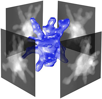
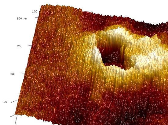
A series of high-resolution scanning transmission electron microscope images of a gold nanostar (four greyscale images), taken from different vantage points, is used to generate a 3D representation of the entire morphology (blue nanostructure at the center).
What is the scientific achievement?
CFN users from Rutgers University worked with CFN staff to perform high-resolution, 3D imaging of metallic nanostructures by scanning transmission electron microscopy (STEM). The measured 3D structure of these nanostars was used as input for finite element simulations of their physical and optical properties, in remarkable agreement with experimental measurements.
Why does this achievement matter?
Nanomaterials can have enhanced optical properties stemming from plasmonic effects, giving them promise for advanced sensors and diagnostic applications. This study represents the first time that information from STEM tomography has been used to predict the physical and optical properties of a nanomaterial.
What are the details?
CFN Capabilities: The CFN Electron Microscopy facility was used to perform characterization by annular STEM tomography.
Gold nanostars are a class of plasmonic nanomaterials that show promise in surface-enhanced Raman scattering (SERS)-based applications and hot electron –driven plasmonic devices. However, important fundamental material properties are difficult to measure because of their complex, spiky morphology—which is fundamental to the field enhancements that make nanostars interesting.
Typically, nanostar properties such as the volume, surface area, and extinction coefficient are merely estimated using a highly simplified, tractable but often inaccurate representation of the morphology. In this work, the authors devised a new method for calculating these fundamental material properties. They used high-resolution, 3D topographic information about individual nanostars as inputs for finite element calculations of the volume, surface area, and morphology-dependent extinction coefficient. Three-dimensional morphologies were obtained by STEM tomography. This new method holds promise for estimating shape-dependent parameters of complex nanostructures of any arbitrary shape and composition—information that is highly relevant for developing plasmonic materials and devices.
Publication Reference
T.V. Tsoulos1, L. Han2, J. Weir1, H.L. Xin2, L. Fabris1
1 Department of Materials Science and Engineering, Rutgers University, Piscataway, New Jersey, USA
2 Center for Functional Nanomaterials, Brookhaven National Laboratory, Upton, New York, USA
Nanoscale 9, 3766 (2017)
Acknowledgement of Support
The work was supported by the National Science Foundation (CHE-1415881). This research used resources of the Center for Functional Nanomaterials, which is a U.S. DOE Office of Science Facility, at Brookhaven National Laboratory under Contract No. DE-SC0012704.
2017-12688 | INT/EXT | Newsroom