NSRL User Guide: Biology Experiments
III. Technical Data
Beam Uniformity and Profile
Before studying beam fragmentation at NSRL, it is important to understand exactly what is the question that is being studied. This discussion is divided into Beam Fragmentation for Physicists and Beam Characterization for Biologists.
Fragmentation for Physicists
The LBL team [1] has conducted an exhaustive program to measure the fragmentation cross sections in heavy ion collisions. They use a pencil-thin beam of ion AB incident on a target ion AT at energy EBeam and look at fragments of the beam ions assuming the fragments are co-moving with the beam, i.e. move at the same velocity as the beam and at zero degrees scattering angle. The fragmentation cross sections have been measured very well down to fragment masses AF equal to half the incident ion mass, (AB)/2. NSRL has continued the work of the LBL team using scintillators to monitor the small angle scattering of beam and target projectiles. Details of these studies are at Fragmentation Physics (pdf).
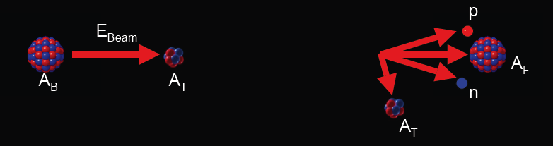
Figure 1 schematic of the fragmentation process showing a heavy ion of atomic weight AB and energy EBeam striking a target nucleus of atomic weight AT. After the collision the projectile has fragmented into a proton, a neutron, and a fragment AF, all of which travel more or less collinearly with the beam. The target particle can also fragment traveling at larger angles from the beam in general.
The fragmentation spectrum measured using a 1000 MeV/nucleon Fe-56 beam shows that fragments with Z anywhere between 1 < Z < 26 are produced. (The peak at Z=25 appears only as a small shoulder on the unfragmented Z=26 peak with well known suppression at Z=4 and 9.)
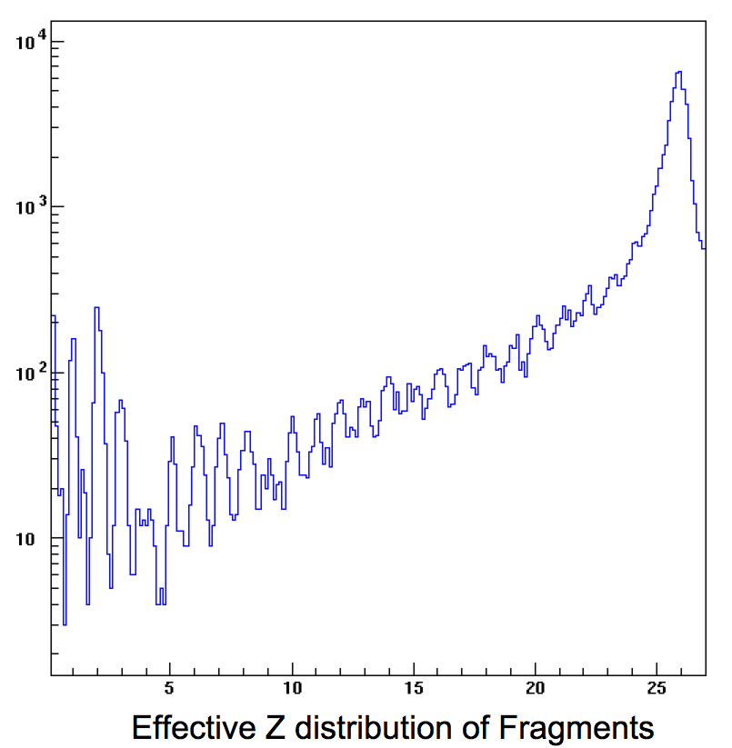
Figure 2 shows the effective Z distribution of the fragments detected in a scintillator placed in a narrow (1 cm diameter) beam of 1000 MeV/nucleon Fe-56.
Measurements like the one above can be used to derive cross sections that are input to Galactic Cosmic Ray simulations.
[1] Zeitlin, Cary et al; Fragmentation of 14-N, 16-O, 20-Ne, and 24-Mg Nuclei at 290 to 1000 MeV/nucleon (2011) Phys. Rev. C. 83. DOI: 10.1103/PhysRevC.83.034909. and Heilbronn, Lawrence et al; Fragmentation Physics and Spacecraft Shielding Studies, (2019) Radioisotopes 68, DOI: 10.3769/radioisotopes.68.407.
Beam Characterization for Biologists
The radiation field seen by biological samples in the beam is not always adequately defined by just the primary heavy ion, even with zero degree fragments included. Sometimes it is important to include all the wide-angle slow-moving low-Z fragments as well. For biological samples, we also measure the composition of the radiation field in and around the NSRL beam.
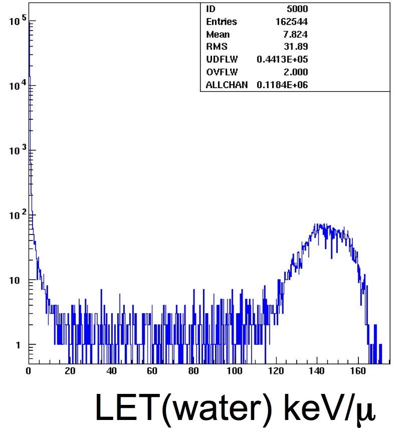
Figure 3 shows the effective LET distribution for all particles incident on samples in the beam. The broad peak near 150 keV/micron is from the Fe-56 ions. The peak at low LET is from protons and low energy photons.
NSRL staff perform these measurements with a selection of scintillators placed directly in the beam, and also at locations in the target room. The scintillators are sensitive to energy loss by charged particles, in much the same ways as biological samples are sensitive to dose; however the scintillator response saturates at high dose rates and needs calibration before the dose-signal response is understood. Details of the effects of fragmentation on the NSRL beam composition are at Beam Composition for Biology (ppt).