NSRL User Guide: Biology Experiments
III. Technical Data
Beam Uniformity and Profile
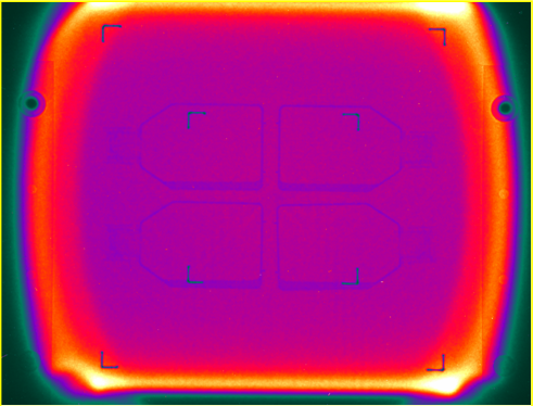
Although the beam profile at NSRL can be tuned to a variety of shapes and sizes, the most common profile used in radiobiology experiments is a "square" beam with a uniform center of approximately 20 x 20 cm2. A picture of this beam profile captured with the Digital Beam Imager is shown below.

Figure 1: Beam Profile observed using the Digital Beam Imager showing four T75 flasks with a small amount of medium in the bottom of each flask. False color indicates the beam intensity is uniform across the 20 x 20 cm2 exposure area. Note that the foam used to hold the flasks is not registered by any change in intensity, and is essentially invisible in this image. Hot spots at the periphery are a by-product of octupole focusing magnets.
The false color image displays relative beam intensity, with black/blue being low intensity and yellow/white being highest. There are fiducial markers on the image plate (corner angle brackets) set at ±5 and ±10 cm from the beam center. The uniformity of color within the central region shows the uniformity of the beam profile. Typical beam uniformities of ±2% are achieved.
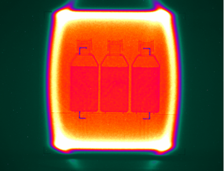
Sometimes smaller beam spots are used in order to achieve higher dose rates or to have the beam entirely contained within the window of an incubator, such as the case illustrated below.

Figure 2: Beam Profile showing three T75 flasks filled with medium. False color indicates the beam intensity is uniform across the 12 x 12 cm2 exposure area. Note that the flasks are supported by a Lucite shelf that can be seen extending across the beam under the flasks.
In addition to large beam spots used for radiobiology, small beam spots can be achieved for high dose work or physics experiments. Although the size of the final focus depends on beam energy and ion species, typical spot sizes of 1 cm RMS can be achieved. Smaller spots or other beam shapes can be produced with the use of collimators. If you have any special demands, contact the NSRL Liaison Physicist (631-344-3072 or 631-344-5830) while you are planning your experiment to ensure that the beam conditions you need will be available.


